
Research Lab
In keeping with the mission of MERC, staff members endeavor to maintain a multidisciplinary research center with core areas of research excellence across a broad spectrum of scientific disciplines.
These include biomechanical testing, computational modeling, histology and histomorphometry, radiography, scanning electron microscopy and particle analysis, in vivo biological modeling, and clinical research.
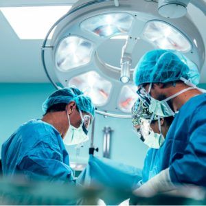
Trained investigators and educational professionals at MERC collaborate with nationally and internationally recognized orthopaedic surgeons and neurosurgeons to conduct research and provide training programs. Our focus is to educate surgeons from all over the world on the safe and effective use of Globus Medical products while providing general education on musculoskeletal disorders and treatment.
Biomechanical testing
The foundation of the biomechanical testing laboratory is the six degrees-of-freedom motion simulator, designed to apply controlled multidirectional motions—flexion/extension, lateral bending, and axial rotation—to musculoskeletal structures. Biomechanical testing platforms are configured with highly accurate optoelectronic motion analysis systems that enable researchers to quantify and compare segmental spinal kinematics following destabilization or reconstruction techniques. The testing platform is designed for a variety of transducers which can be interfaced to measure parameters such as intervertebral disc pressure and strain within osseous structures or spinal constructs.
The laboratory is also equipped with MTS units for static and fatigue testing of specimens, a dual-energy x-ray (DEXA) scanner for quantification of bone mineral density, an O-arm surgical navigation system, and fluoroscopic image intensifiers.
Computational Modeling
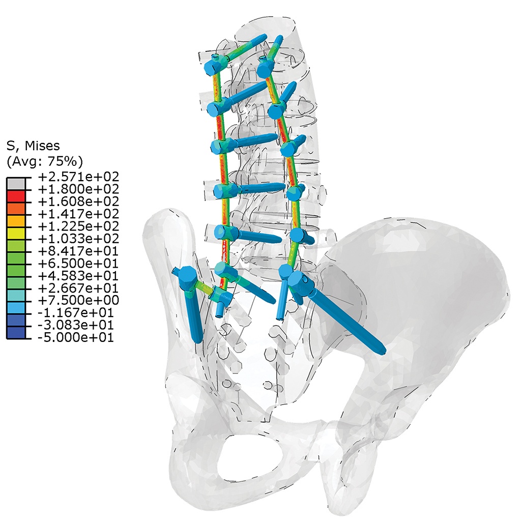
Along with biomechanical testing equipment, the MERC facility features a computer-aided design and computational analysis software suite. Researchers conduct stress analysis simulations of the spine and applied implant constructs as part of the research and development process.
Finite element models of fully anatomic L4-L5 lumbar motion segments and lumbopelvic anatomies generate accurate, experimentally validated data sets. These models allow physiologic simulation of standard decompression, disc replacement, and fusion reconstruction techniques, and enable researchers to investigate stress and strain patterns within implants or biological structures. Computational modeling provides the opportunity for surgeons and engineers to improve implants designed for fusion or non-fusion technologies, streamline the product development life cycle, and conduct exploratory analysis of the anatomy.
Histology and Histomorphometry
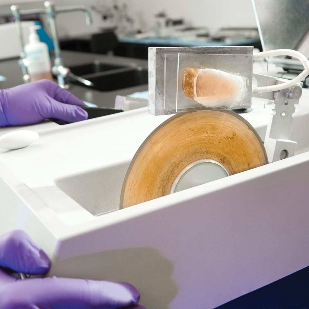
The histology laboratory is capable of processing a wide variety of tissue samples that include operative fusion segments, intervertebral arthroplasty devices, osseous defects, and implants, through undecalcified and decalcified techniques. The laboratory is uniquely equipped for examination of oversized osseous specimens using micro-grinding technology, microtomes, and diamond cutting blades.
Histologic slides of varied thickness can be produced for highly accurate assessment of bone remodeling, wear debris, inflammatory reactions, degenerative changes, and osseointegration at the implant interface. Immunohistochemical macrophage and cytokine assays can be performed to further delineate cellular mediators in inflammatory reactions. Histomorphometric image analysis permits quantitative calculations of osseous and soft tissue parameters.
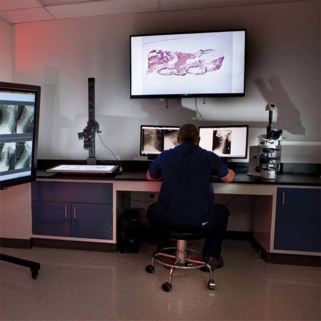
Radiography
A comprehensive suite dedicated to visualization and quantitative assessment of radiographic plain films, high resolution microradiographs, computed tomography (CT), micro computed tomography (MicroCT) scans, and magnetic resonance imaging (MRI), is available. The MicroCT core facility houses a high-energy desktop scanner with the flexibility to image large and small objects of variable density. MicroCT offers greatly enhanced resolution, which approaches tens of microns, and provides a method of conducting 3D microscopy that can be applied in analysis of spinal implants and biological materials. Biological tissue samples can be imaged in combination with implants and virtually delineated with the use of image registration and segmentation software. Moreover, 3D slicing algorithms can be used to quantify bone volume in multiple planes and orientations as observed on CT/MRI and MicroCT scans. Applications include measurement of bone formation and implant resorption, quantification of bone volume, and overall morphologic assessment.
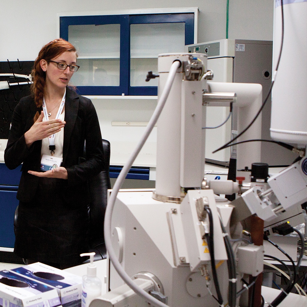
Scanning Electron Microscopy and Particle Analysis
The scanning electron microscopy and particle analysis laboratory at MERC permits researchers to evaluate implant surface topography and perform particle analysis—processes that are paramount for fusion and non-fusion technologies. The scanning electron microscope (SEM) is capable of analyzing wear particulates and surface topography at magnifications greater than 100,000. The SEM can be used in combination with computational modeling techniques to determine how corrosion, pitting, surface erosion, cracks, and particulate are related to stress under in vivo conditions. Results of surface particulate analysis can be combined with SEM topographic images for complete characterization of the surface environment. Energy dispersive x-ray spectroscopy can be performed to identify unknown materials by elemental composition.
In Vivo Biological Modeling
MERC conducts a wide variety of pre-clinical in vivo modeling studies to assess safety and efficacy of implant materials and devices. Strict adherence to all applicable guidelines as outlined by the Code of Federal Regulations (CFR, Title 21, part 58) and the USDA Animal Welfare Act is of utmost importance in these studies. All facilities interacting with MERC are accredited by the Association for Assessment and Accreditation of Laboratory Animal Care, compliant with the National Institutes of Health Office of Laboratory Animal Welfare, licensed by the US Department of Agriculture, and inspected by the Food and Drug Administration.
Research projects are approved and closely monitored by the Institutional Animal Care and Use Committee, and strictly adhere to all applicable stipulations of the most recent version of the USDA Animal Welfare Act and Good Laboratory Practices. MERC has extensive experience with in vivo biological modeling for spinal arthrodesis, bone graft substitutes, motion preservation–based surgical reconstruction techniques, and neurotoxicity models.